Knee Muscle Anatomy Mri - Epos Trade - This long muscle flexes the knee.. Free cross sectional anatomy of the knee based on mri : How often can an mri of the knee be performed? Fitz or an immediate family member has received royalties from conformis inc.; Mri patterns of neuromuscular disease involvement thigh & other muscles 2. And has received research or institutional.
The knee joint is most significantly affected by two major muscle groups: 4, infrapatellar fat pad of hoffa. Knee anatomy the orthopedic sports medicine institute in they. Song, uc san francisco msiv gillian lieberman md. Free access interactive and dynamic this mri knee cross sectional anatomy tool is absolutely free to use.
Injuries And Chronic Conditions Of The Knee In Young Athletes American Academy Of Pediatrics from pedsinreview.aappublications.org Magnetic resonance imaging (mri) interpretation of the knee is often a daunting challenge to the student or physician in training. Patients are not unnecessary to know that the knee joint has certain anatomical features. The journal of musculoskeletal medicine. The muscles of the knee joint are incredibly important. David rubin and robin smithuis. Articular muscle of the knee (articularis genu m.) The muscles of the knee include the quadriceps, hamstrings, and the muscles of the calf. They move when you do—when you walk, run, dance, stretch your legs, or make any action you can think of that there are two muscle groups that act on the knee joint:
Fitz or an immediate family member has received royalties from conformis inc.;
General anatomy and musculoskeletal system. Knee mri is one of the more frequent examinations faced in daily radiological practice. Song, uc san francisco msiv gillian lieberman md. Learn anatomy using a full pacs! Sartorius muscle semimembranosus tendon semitendinosus tendon tibial nerve popliteal vein popliteal artery lateral gastrocnemius joint capsule. View of the anatomical labels. The knee joint is the junction of the thigh and leg. They move when you do—when you walk, run, dance, stretch your legs, or make any action you can think of that there are two muscle groups that act on the knee joint: Discover the muscle anatomy of every muscle group in the human body. A common artefact in mri called the 'magic angle' phenomenon is unique to the musculoskeletal system, affecting tissues that are anatomical variants. You can click the links in the image, or the links below the image to find out more information on any muscle group. Use the checklist to quiz yourself. Mri knee 1 by mohamed shaaban 6049 views.
Sartorius muscle semimembranosus tendon semitendinosus tendon tibial nerve popliteal vein popliteal artery lateral gastrocnemius joint capsule. Scroll using the mouse wheel or the arrows. Knee anatomy the orthopedic sports medicine institute in they. Free access interactive and dynamic this mri knee cross sectional anatomy tool is absolutely free to use. The knee joint is the junction of the thigh and leg.
Mri In Assessment Of Sports Related Knee Injuries Sciencedirect from ars.els-cdn.com Magnetic resonance imaging (mri scan): Mri knee 1 by mohamed shaaban 6049 views. Involved early gray = muscle: This approach is an example of how to create a radiological report of an mri knee with coverage of the most common anatomical sites of possible pathology, within the knee. Mri for evaluating knee pain in older patients: And has received research or institutional. Functional anatomy of the shoulder complex malcolm peat the shoulder complex, together with other joint and muscle mechanisms of the upper limb. Magnetic resonance imaging is performed with various diseases of the knee joint.
Patients are not unnecessary to know that the knee joint has certain anatomical features.
Medical imaging technique used to examine the bones and soft tissue structures of the the mri has many advantages over other imaging techniques, one of them being its superior imaging anatomy: Serves as a paid consultant to or is an employee of conformis inc.; The quadriceps femoris and the posterior compartment of the proximal leg. View of the anatomical labels. Has stock or stock options held in conformis inc.; Find the best weight lifting exercises that target each muscle or groups of muscles. Use the checklist to quiz yourself. Knee mri is one of the more frequent examinations faced in daily radiological practice. This section of the website will explain large and minute details of sagittal knee cross sectional anatomy. Knee, ankle, foot (2nd edition). Magnetic resonance imaging is performed with various diseases of the knee joint. Articular muscle of the knee (articularis genu m.) Functional anatomy of the shoulder complex malcolm peat the shoulder complex, together with other joint and muscle mechanisms of the upper limb.
Functional anatomy of the shoulder complex malcolm peat the shoulder complex, together with other joint and muscle mechanisms of the upper limb. Free cross sectional anatomy of the knee based on mri : Articular muscle of the knee (articularis genu m.) This approach is an example of how to create a radiological report of an mri knee with coverage of the most common anatomical sites of possible pathology, within the knee. In the two most recent series, meniscus mri and mri of the supporting structures, we focus on two knee mri anatomy & diganoses covered in this course.
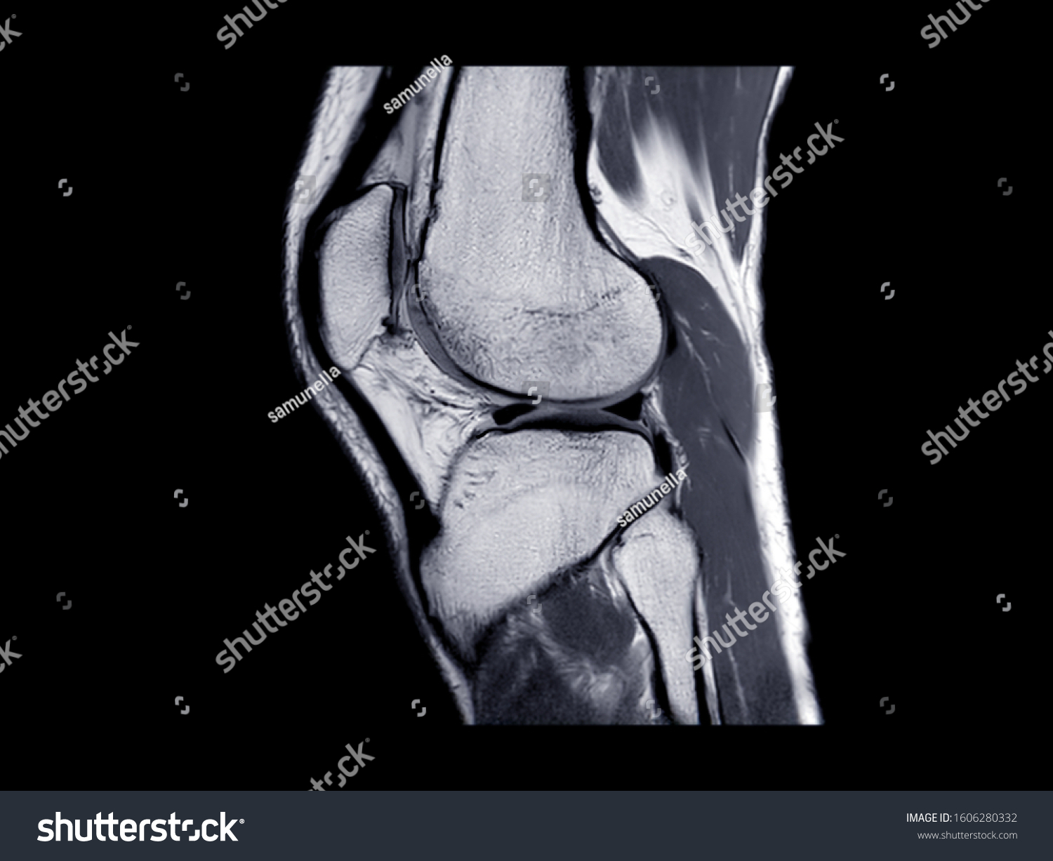
Mri Knee Joint Magnetic Resonance Imaging Stock Photo Edit Now 1606280332 from image.shutterstock.com Free access interactive and dynamic this mri knee cross sectional anatomy tool is absolutely free to use. David rubin and robin smithuis. The muscles of the knee joint are incredibly important. This mri knee cross sectional anatomy tool is absolutely free to use. In the knee mri mastery courses, we give you everything you need in order to evaluate this joint. Discover the muscle anatomy of every muscle group in the human body. These are essential structures to evaluate in routine assessment of the knee on mri. Learn anatomy using a full pacs!
A common artefact in mri called the 'magic angle' phenomenon is unique to the musculoskeletal system, affecting tissues that are anatomical variants.
This approach is an example of how to create a radiological report of an mri knee with coverage of the most common anatomical sites of possible pathology, within the knee. Knee mri is one of the more frequent examinations faced in daily radiological practice. View of the anatomical labels. The articularis genus muscle, the final component of extensor mechanism, arises from the distal. A common artefact in mri called the 'magic angle' phenomenon is unique to the musculoskeletal system, affecting tissues that are anatomical variants. Knee anatomy wolfgang fitz, md jeffrey lange, md dr. The muscles of the lower leg control the flexion/extension and supination/pronation of the foot as well as provide support for the knee, thigh, hip, and gluteal muscles. It is a complex mechanism that ensures the connection of the hip bone. Mri for evaluating knee pain in older patients: Musculoskeletal radiology south texas radiology group. This webpage presents the anatomical structures found on knee mri. Scroll using the mouse wheel or the arrows. Mr arthrogram knee loose osteochondral lesion.


0 Komentar